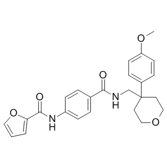
This is consistent with the concept that once proteins are transported into the proteasome, they cannot exit until degraded and if the preferred enzyme is not active, then cleavage by the other subunits becomes the primary route of degradation. Another previous study examined the effect of bortezomib on the cellular peptidome. Bortezomib is a reversible proteasome inhibitor containing an active site boronate group and is FDAapproved to treat multiple myeloma and mantle cell lymphoma. Bortezomib is a potent inhibitor of the beta 5 subunit, and at higher concentrations blocks the beta 1 subunit. Because the beta 5 subunit plays a major role in the conversion of proteins into peptides, and bortezomib potently inhibits this subunit, it was expected that this drug would cause a decrease in the levels of these peptides, as found for epoxomicin. However, the opposite result was found; the majority of intracellular peptides was elevated by treatment with bortezomib, including many peptides that were predicted to be products of beta 5 cleavages. One possible explanation of this paradoxical result is that bortezomib has off-target effects on the enzymes that degrade the intracellular peptides; a previous study predicted that bortezomib may inhibit TPP2, based on the finding that bortezomib inhibited other cellular serine proteases such as cathepsins A and G. Alternatively, bortezomib is known to allosterically influence proteasome stability, gate opening, and cleavage specificity, and it is possible that these allosteric effects cause the increase in cellular peptides upon exposure to bortezomib. To study this, we used a peptidomics method to examine the effect of a variety of proteasome inhibitors on the peptidome of HEK293T and SH-SY5Y cells; these cell lines were used because their peptidomes have been well-studied. The inhibitors picked for this analysis include three boronate compounds that inhibit the proteasome reversibly, and three non-boronate compounds, one of which is an irreversible inhibitor and two of which are reversible inhibitors. Carfilzomib is an analog of epoxomicin that was recently approved for the treatment of multiple myeloma and mantle cell lymphoma. Some of these proteasome inhibitors are known to have off-target effects, such as MG132 which inhibits calpain and clasto-Lactacystin b-lactone which inhibits cathepsin A. We also tested bortezomib as an inhibitor of peptidases present in HEK293T cells using assays that detect TPP2 and puromycinsensitive aminopeptidase. Finally, we examined whether potent inhibitors of these two enzymes Niltubacin HDAC inhibitor influenced the peptidome of HEK293T cells. Although bortezomib, MG262, and one of the other boronate-containing proteasome inhibitors are weak inhibitors of HEK293T cell aminopeptidase activity, this effect does not appear to contribute to the large increase in most cellular peptides observed with bortezomib and MG262, and to a lesser extent, with carfilzomib. To identify peptides, MS/MS data were analyzed using the Mascot search  engine and the IPI_human data base. LEE011 Searches include variable modifications of N-terminal acetylation, methionine oxidation, and the isotopic D0-, D3-, D6-, and D9TMAB tags used in our study. Results were manually interpreted to eliminate false positives, using previously described criteria. To investigate the discrepancy between the effect of epoxomicin and bortezomib on the levels of intracellular peptides, we tested six additional proteasome inhibitors. Because AM114 contains two boronate groups, it was included in subsequent studies to test if the previously observed effects of bortezomib were caused by off-target effects due to the boronate groups rather than inhibition of the proteasome.
Category: GPCR Compound Library
The importance of circulating endothelial progenitor cell number measured as CD34
Our study now provides evidence that in vivo and in vitro the number of VPC is reduced in conditions of type 2 diabetes and that one major signalling pathway involved is the activation of the transcription factors ETS1 and ETS2. The ETS protein family is a group of transcription factors that share a DNA-binding ETS domain and regulate the expression of a variety of genes including key ones involved in regulation of cell proliferation, differentiation and survival. Because of their critical role in basic cellular processes, dysregulation of ETS transcription factors can be found in many human diseases with the need for neovascularisation, such as cancer. ETS transcription factors have been implicated in the regulation of genes involved in homeostasis, vascular development and angiogenesis, even though no endothelial specific ETS transcription factor has been identified by now. We could clearly show that ETS dysregulation by high levels of glucose leads to impairment of progenitor cell number as well as functional capacity. Apoptozole KDR + cells in the peripheral blood, has recently been shown as these cells predict the outcome in CAD-patients. Although the transcription factor ETS reduces VPC numbers, ETS activity is clearly important in adult angiogenesis. In the resting endothelium, ETS1 is expressed at a very low level. During angiogenesis, re-endothelialisation after balloon denudation in a rat model as well as in scratch wound migration assays, ETS1 is transiently expressed at high levels in endothelial cells, suggesting that during the process of vessel formation or repair upregulation of ETS1 transcription factor expression is required in mature endothelial cells. However, the process of preexisting mature endothelial cells to perform angiogenesis differs completely from VPC-induced vasculogenesis or vascular repair of the damaged endothelial layer. Even though our findings demonstrate the importance of ETS1 in angiogenesis, ETS1 deficient mice develop normally except for an increased perinatal mortality. Especially no vascular phenotype can be detected. Therefore, we hypothesized that most likely other ETS transcription factors can compensate for its loss. Alternatively, these mice might have increased numbers and improved functionality of their endothelial progenitor cells for compensation. Members of the ETS gene family are known to be expressed in the hematopoietic tissue and some of them play a pivotal role in hematopoietic cell development. The special importance of ETS1 regulation in differentiation of Importazole hematopoietic stem cells has previously been shown. During erythroid differentiation, ETS1 is downregulated and exported out of the nucleus. In contrast, during megakaryopoiesis, ETS1 increases and remains in the nucleus. Similarly, it seems that ETS1 activation is required for angiogenesis by sprouting of mature endothelial cells and down-regulation of ETS DNA binding activity is required for increased VPC differentiation from their progenitors as shown in this study. We clearly showed that glucose upregulates ETS DNA binding activity and thereby reduces VPC numbers. In addition, it has been shown that other pro-inflammatory stimuli/cardiovascular risk factors such as TNFa up-regulate ETS transcription factors and reduce the number of endothelial progenitor cells. Therefore, it is tempting to speculate that not only high glucose but also TNFa prevents endothelial cell lineage commitment by ETS transcriptions factor activation.
The expression levels of Tlr4 mRNA in LPS/GalN induced hepatic injury were significantly increased compared to the control
It has also been shown that neutralizing antibodies to HMGB1 amelliorate liver I/R injury, thereby suggesting a therapeutic benefit of blocking active HMGB1 release to minimize I/R-associated damage.In contrast, our preliminary work presented the results that the intravenous injection of HMGB1 6 hours after LPS/GalN-treatment or HMGB1 alone did not precipitate ALT/AST activity. Furthermore, the intravenous injection of neutralizing antibodies to HMGB1 did not ameliorate an increase in serum ALT/AST activity in LPS/GalN-injured mice. In addition to these findings, but those of Rage mRNA conversely decreased in this hepatic injury. Considering together with these data, acute hepatic injury stimulated by a single injection of LPS/GalN might not be caused by HMGB1 released into the extracellular milieu from non-hematopoietic cells or activated macrophages/dendritic cells. In the current investigation,SAR405 numerous apoptotic cells were observed in the pericentral areas of LPS/GalN-treated liver. To understand mechanisms of hepatocyte apoptosis promoted in the LPS/GalN-induced hepatic injury, we explored the fluctuation in the expression of genes associated with the regulation of apoptotic cell death using the gene microarray analysis. Mouse GSTO1 was indeed identified because of its overexpression in a mouse lymphoma cell line presenting with resistance against a variety of chemo-therapeutics. After transfection with Gsto1 siRNA, transfected HeLa cells present with a marked increase of apoptosis as compared with controls, suggesting that GSTO1 plays an anti-apoptotic role. GSTO1 overexpression is associated with activation of survival pathways including Akt kinase and extracellular signal-regulated kinase 1/ 2 and anti-apoptotic pathways of c-Jun N-terminal kinase 1. Immunohistochemistry of LPS-treated liver remnants showed the remarkable decrease of nuclear reactivity and sparse reactivity of cytoplasm with the antibody to activated p65 at 8 or 10 h compared with controls or GL-treated remnants. Although the activation of NF-kB is associated with potent inflammatory responses in hepatic injury, key roles for this factor in inhibition of apoptosis in the liver have been also demonstrated definitively experimentally. In conclusion, G007-LK in mouse liver injury induced by a single injection of LPS/GalN, HMGB1 protein appears to be implicated in the inhibition of a signal pathway associated with anti-apoptosis, i.e. down-regulation of GSTO1, and to stimulate apoptosis of hepatocytes rather than to act as pro-inflammatory factors in this experimental model. However, we cannot exclude the possibility that additional LPS-treatment induces release of HMGB1 into extracellular milieu and precipitates hepatic inflammation through the receptors of TLR4 and/or RAGE. In addition to the role of GL disclosed in this study, it has been reported that GL treatment inhibits the proliferation and migration of cells stimulated by HMGB1 cytokine, as well as HMGB1-induced formation of blood vessels and reduces inflammatory condition. There is an on-going need to develop new agents and provide new strategies for the treatment of hospital- and communityacquired infections caused by Staphylococcus aureus. This opportunistic pathogen has proven to be adept at acquiring genes encoding resistance mechanisms against front-line antibiotics and, in the absence of appropriate preventative measures, multi-drugresistant forms such as methicillin-resistant S. aureus clones are able to disseminate at sometimes alarming rates amongst patients in healthcare facilities.
PLB-SERCA2a system could be an upstream regulator of miR-21 in the b-adrenergic signaling cascade
MiR-21 is overexpressed in many tumors and considered as key regulator of oncogenic processes. It is also highly expressed in CM and cardiac fibroblasts and apparently plays an important role in cardiac metabolism. Both miR-21 regulation as well as PLB-SERCA2a interaction are known to be regulated via b-adrenergic receptor signaling. In the dairy cow, late gestation and early lactation are periods marked by major changes in the sensitivity and responses of tissues to hormones involved in homeostasis, such as insulin. Indeed, during these periods, there is a moderate decrease in peripheral tissue insulin sensitivity,HDM201 promoting the mobilization of non esterified fatty acids and amino acids and facilitating the preferential use of nutrients by the fetus or mammary gland. The decrease in insulin sensitivity occurring in adipocytes during late gestation and early lactation in dairy cows remains poorly understood. Insulin acts by binding to the insulin receptor, a tyrosine kinase receptor, on cells. Following insulin binding, the IR phosphorylates various substrates, including IRS-1 and IRS-2, which interact with several intracellular proteins to activate different signaling pathways, including the PI3K/Akt and MAPK ERK1/2 pathways. Sadri et al. studied the expression of genes encoding components of the insulin receptor signaling pathway in adipose tissue during the dry period and in early lactation, in dairy cows. They observed a significant decrease in insulinresponsive glucose transporter gene expression in subcutaneous adipose tissue around the time of parturition. However, the levels of phosphorylation of IR signaling components have never been investigated. Adipokines factors secreted by the adipose tissue may be involved. In particular, resistin is known to decrease insulin sensitivity in rodents, whereas its effect in humans is unclear. Resistin is a protein consisting of 108 amino acids in humans, 114 amino acids in mice, and 109 amino acids in cattle; it belongs to the “resistin-like molecules” or “FIZZ” family. It consists of homodimers linked by disulfide bridges. Resistin is produced directly by the adipocytes in mice, whereas it is produced by macrophages and transported to adipocytes in humans. Plasma resistin levels are correlated with the degree of insulin resistance in mice,BMS-935177 whereas conflicting results have been reported concerning this aspect in humans. In bovine species, the localization in adipose tissue and the role of resistin in lipolysis are still unknown. In mice, plasma and adipose tissue levels of resistin decrease in response to thiazolidinediones and increase during obesity. Very little is currently known about the mode of action of resistin. No receptor has yet been clearly identified and the signaling pathways used remain unclear. Recent studies have suggested that resistin may bind a receptor tyrosine kinase called ROR1 in murine pre-3T3-L1 adipocytes, or to TLR4 in the hypothalamus of mice. The adipose tissue of dairy cows also produces several adipokines, including resistin. Komatsu et al. showed that levels of resistin gene expression in the adipose tissue were significantly higher in lactating than in non lactating cows, whereas the opposite pattern was observed in the mammary gland.
Determining whether CK5 mRNA levels can be linked to the development of either acute or chronic rejection
By implication, it would appear that circulating epithelial progenitor cells most likely do play an important role in airway repair, although whether this is a direct of indirect effect still needs to be established. Ultimately, infection is critical and is an area of active investigation in the lab. In summary, CK5 expressing circulating epithelial progenitor cells are present in circulation in normal human subjects and can be quantified with the real time PCR assay that we have established. Furthermore,Brivudine the circulating epithelial progenitor cells are significantly reduced immediately after lung transplantation, and then increase with time as lung function improves. We suggest that circulating epithelial progenitor cells may play an important role in airway repair after lung transplantation. In addition, circulating epithelial progenitor cells may be used as a biomarker of airway repair. Cocaine is a plant alkaloid derived from coca plant leaves and represents a major drug of abuse. In animal models, acute administration of cocaine evokes changes in locomotor activity, grooming, and feeding, and can induce uncontrolled repetitive behaviors. At the cellular level, cocaine elevates extracellular monoamine levels by inhibiting monoamine reuptake transporters, including DAT, SERT and NET. By acting on their cognate receptors, monoamines elicit both short-term and long-lasting alterations in the nervous system, which ultimately lead to the development of drug dependence. While dopamine is generally believed to be a principal neurotransmitter functioning in the mesolimbic dopamine system to mediate drug dependence, ample evidence suggests that other neurotransmitter systems are also required for the expression of drug addiction behaviors. In particular,Cefotiam hydrochloride serotonin is believed to play an important role in mediating the reinforcing effects of cocaine. For example, the induction of conditioned place preference by cocaine is normal in DAT knockout mice, but is eliminated in mice lacking both DAT and SERT. Further, it has been shown that that DAT knockout mice can still self-administer cocaine, though a recent study has challenged this finding. Therefore, to better understand the mechanistic underpinnings of drug addiction, and to develop new therapeutic interventions, a greater knowledge of the genes and molecules regulating cocaine’s behavioral effects is required. Despite their simplicity, invertebrate model organisms such as C. elegans and Drosophila are widely used in neurobiology and have yielded novel insights into relatively complex behavioral phenomena, including drug dependence. Furthermore, the major genes found to be involved in drug dependence are conserved in these organisms. The powerful genetics of invertebrate models, combined with their short generation time, make these organisms a valuable resource for the study of basic mechanisms underlying drug-induced behaviors. In the present study, we tested the effect of cocaine on C. elegans locomotion behavior. We find that acute cocaine treatment alters its locomotor activity. This behavioral response to cocaine is mediated by serotonin. In this study, we have shown that C. elegans responds to acute cocaine treatment by reducing locomotor activity, a behavioral response that is mediated by the neurotransmitter serotonin.