Hence, functional analyses will ultimately be required to definitively appreciate the mechanisms of TLR5/flagellin dimerization. Our results would indicate that on the whole, greater functional activity of the TLR5 variant rs5744174 is a risk factor for CD in children. Reasons for this remain speculative, but could include differences in recruitment of Th17 cells or dendritic cells to the intestinal lamina propria, leading to altered inflammatory tone. Our conclusions are limited by the need to use a non-intestinal cell line for functional studies, since human intestinal epithelial cells and leukocytes express native TLR5. Testing of a large cohort of volunteers with both SNPs of TLR5 at residue 616 would be required to draw conclusions about functional differences in flagellin response, because of naturally occurring polymorphisms in other inflammatory genes involved in chemokine and cytokine production. However, these future important predict precisely prognosis therapeutic effect studies could provide interesting information to guide new diagnostic or therapeutic interventions for pediatric CD. It is a public health threat due to the potentially disastrous results and high cumulative rate of fractures. It is estimated that more than  100 million people worldwide are at risk for the disorder and fracture rates seem to be rising ceaselessly. Osteoporosis is mainly caused by an imbalance between osteoblast-mediated bone formation and osteoclast-mediated bone resorption. A number of medicines have been developed to treat osteoporosis, mainly including bone resorption inhibitors, which prevent excessive bone loss by reducing the osteoclast formation and activity; bone formation accelerators, which increase bone mineral density and bone mass by stimulating the osteoblast activity; bone mineralization drugs, which stimulate new bone mineralization. NUCB21�C83 is also called nesfatin-1. It has recently been identified as a satiety molecule associated with melanocortin signaling system detectable in central neurons as well as an anti-hyperglycemic peptide when it is given intravenously. NUCB21�C83 was also reported to have a role in the response to stress and mediation of anxiety- and/or fear-related behaviors in rats. The expression of NUCB21�C83 was induced by troglitazone, an activator of peroxisome proliferator-activated receptor-c. The activation of PPAR-c was recognized to cause loss of bone. Therefore, we have curiously examined the effect of NUCB1�C83 on bone metabolism. Since ovariectomized rat is a classic animal model for postmenopausal osteoporosis, we have intravenously injected NUCB21�C83 once a day to OVX rats continuously for two months to observe the changes in bone mineral density. In addition, we have also evaluated both the promoting effect of NUCB21�C83 on osteoblastogenesis in the mouse MC3T3-E1 preosteoblastic cell line and its inhibitory effect on osteoclastogenesis in murine RAW 264.7 macrophages, as well as its presence in osteoblasts and osteoclasts. The nucleobindins, NUCB1 and NUCB2, are homologous calcium and DNA binding proteins. It was reported that secreted extracellular NUCB1 might contribute in modulating the matrix maturation in bone with unknown mechanisms. NUCB21�C83 was originally identified as an anorexigenic factor in hypothalamus which was recently reported to be anti-hyperglycemic. However, it has not been reported to affect bone metabolism. In our experiments, the intravenous administration of NUCB21�C83 was found for the first time to increase BMD of femora and lumbar vertebrae in OVX rats. Mouse MC3T3-E1 preosteoblastic cell line was derived from calvaria of newborn mice. It has been widely applied in the investigation of mechanism underlying osteoblast differentiation as it epitomizes osteogenic differentiation and maturation in vitro. ALP is a representative marker for osteoblastic differentiation.
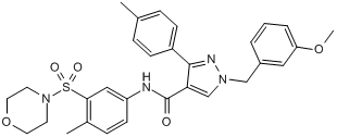
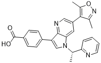
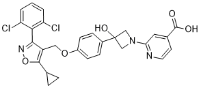
Category: GPCR Compound Library
For the development of premature atherosclerosis in SLE patients
Therefore, there is an interest in using anti-inflammatory or edna techniques efficient inventory monitoring programs sensitive species immunomodulatory therapies for this condition. MMF, an immunosuppressive agent, is currently used for the treatment of SLE patients, particularly those with kidney involvement, as well as for the prevention of rejection in transplant patients. MMF was first demonstrated to inhibit T-cell function, however, MMF also exerts inhibitory effects on other immune cells and effectors, including downregulation of cell adhesion molecules and attenuation of monocyte and macrophage responses. MMF has also been shown to suppress a number of the inflammatory events that are involved in the development of atherosclerosis. T-lymphocyte infiltration to atherosclerotic plaque and circulation  to sites of inflammation is abrogated by MMF treatment. MMF has been shown to reduce the expression of vascular adhesion molecules in atherosclerosis by inhibiting the nuclear factor NFkB which is required for their transcriptional upregulation. Raisanen et al showed that MMF treatment reduces the appearance and proliferation of smooth muscle cells in the intima, which normally contribute to atherosclerotic plaque formation by recruitment of extracellular matrix and self-proliferation. Thus, MMF has properties that could be considered anti-atherogenic: inhibiting T-cells, blocking leukocyte adhesion and inhibiting proliferation of smooth muscle cells; therefore making it a potentially valuable drug to prevent the development of atherosclerosis in patients with SLE. A recent publication examined the effect of MMF on atherosclerosis development by using bone marrow transplantation to generate a mouse model of lupus with associated atherosclerosis. Substantial evidence exists to support a critical role of inflammation in the pathology of both SLE and CVD caused by atherosclerosis. The chronic inflammation associated with SLE correlates to the increased risk in CVD seen in patients. The current study suggests that MMF, in addition to this wellknown efficacy on lupus nephritis, might be a promising agent for the prevention of atherosclerosis in SLE. A more extensive analysis of atherosclerosis and SLE in these MMF-treated gld.apoE2/2 mice will need to be performed to fully determine how MMF impacts the accelerated atherosclerosis of our lupus mouse model and potentially bring a better understanding of the link between atherogenesis and autoimmune disease. The worldwide incidence of obesity has increased dramatically during recent decades. The “thrifty genes”, which were historically advantageous, may now be detrimental due to the abundance of the food supply and dramatic changes in modern lifestyle, and contribute to the major epidemic of obesity. Obesity is associated with a high incidence of steatosis, insulin resistance and chronic inflammation. Obesity-related non-alcoholic fatty liver disease has recently been recognized as one of the major causes of chronic liver disorders, estimated to affect at least one-quarter of the general population. NAFLD is characterized by excess liver lipid accumulation, hepatic insulin resistance, and later hepatic inflammation, leading to nonalcoholic steatohepatitis and culminating in hepatic fibrosis or cirrhosis. One of the major causes of fat accumulation in NAFLD is the inability of the liver to regulate the changes in lipogenesis in the transition from fasted to fed state. Several studies suggested that hepatic lipogenesis is increased in hepatic steatosis, which may result from either increased triglyceride synthesis or decreased fatty acid oxidation through production of malonyl- CoA, both leading to increased triglyceride content in the liver. Excess fat accumulation ultimately leads to hepatic steatosis and worsening hepatic insulin resistance via a network of transcription factors, which regulate hepatic lipogenesis and fatty acid oxidation.
Alternatively a laforin sample purified and stored in the presence of DTT was fully active
While plants lack a true laforin ortholog, laforin is conserved in the genome of five protozoans. We previously demonstrated that Cm-laforin possesses the same biochemical signature as laforin, in that they both bind glucans and can dephosphorylate phospho-glucans. Given the functional similarities between human laforin, Cm-laforin, and SEX4, we used SEX4 and Cm-laforin in the current study to evaluate if the oligomerization phenomenon is true of all glucan phosphatases. We purified SEX4 and Cm-laforin using a similar two-step purification method that included size exclusion chromatography. For each protein, we observed that multiple peaks were eluted from the column, similar as we observed for human laforin. We analyzed the ratio of SEX4 and Cm-laforin monomer and dimer fractions via immunoblotting, as was performed for Hs-laforin in Figure 2B. Then we tested the phosphatase activity of SEX4 using both pNPP and malachite green assays. We found that monomeric SEX4 has a slightly higher specific activity than dimer using both pNPP and malachite green as substrates. For Cm-laforin, the specific activity against pNPP of the monomer form was three times higher than that of dimer; whereas the malachite green assay showed that the glucan phosphatase activity of Cm-laforin was similar for monomer and dimer. Hs-laforin, SEX4, and Cm-laforin all belong to glucan phosphatase family. These results demonstrate that glucan phosphatases from different Kingdoms are all active in the monomeric form. Interestingly, dimeric forms of glucan phosphatases from different Kingdoms exhibit different phosphatase activity, suggesting different modes of actions for each dimeric form. The finding that storage of laforin in low levels of DTT is necessary prompted us to further examine the effect of reducing agents on laforin oligomerization and phosphatase activity. When we analyzed the non-reducing peak of laforin by gel electrophoresis under non-reducing conditions, we observed the presence of laforin monomers, dimers, and multimers. However, if we added increasing amounts of DTT we found that laforin oligomerization was reversed and at 100 mM DTT only monomeric laforin remained. These results suggest that laforin oligomerization is very sensitive to oxidation, and that multiple species of laforin form under non-reducing conditions. These species may result from intermolecular disulphide bond formation among the nine cysteine residues present in laforin. Additionally, these results show that the amount of DTT commonly utilized in phosphatase assays does not affect dimerization or multimerization. However, these low levels of DTT are necessary to keep the catalytic cysteine reduced. Dual specificity phosphatases employ a two-step catalytic mechanism. After nucleophilic attack of the substrate phosphorus atom, a phosphoryl-cysteine intermediate is formed before hydrolysis of the intermediate and release of phosphate. In oxidative environments, this catalytic cysteine is modified and inactivated. In order to define the relationship between oxidation of laforin and its phosphatase activity, we examined the phosphatase activity of laforin that was purified and stored in the absence of DTT using the exogenous substrate 3-O-methyl fluorescein phosphate. We found that the  phosphatase activity of laforin was dependent on the presence of DTT in the reaction buffer: without DTT the activity was abolished, whereas in the presence of 10 mM DTT the activity was significantly higher.
This has actually been one more in-depth and also exact topic on Variability of MDD and CRHR1 gene is likely to be involved in the antidepressant response in MDD, to learn even more and sustain our job, please see http://www.tolllikereceptor.com/index.php/2019/02/24/striking-lead-improvement-lv-ef-long-term/.
In addition to standard computational stereotypic movements, weight loss and cognitive abnormalities
As in the human disease, CAG repeat lengths appear to be associated with disease onset and severity. However, mice with extreme repeat lengths in this model present with a disease having a delayed phenotype. This delay in the onset and reduction in the severity of symptoms, in parallel with neurodegenerative changes, provides a model with the potential to elucidate more of the underlying pathogenesis. In addition to the R6/2 model, a number of other models have been made that were aimed at recapitulating better the genetics of HD. These include full length knock-in models, a yeast artificial chromosome model, and a bacterial artificial chromosome model. Historically, magnetic resonance imaging findings for individual patients were diagnostic only in later stages of HD, for example where caudate atrophy contributed to the characteristically large ventricles seen. More recently, analytical methodologies, such as tensor-based morphometry, have been used to show progressive structural changes in presymptomatic HD patients. Neuroimaging studies based on voxel-based morphometry are also used widely to investigate developing pathology in humans and assess prospective treatments. These automated methods for characterising structural differences or changes in the living brain have also been used in mouse models to show that many pathological features are shared between the mouse models and humans with the disease. Here we describe a large dataset of MR images of mice used in models of HD that includes transgenic R6/2 lines of various CAG expansion lengths, yeast artificial chromosome and wildtype mice. In addition, we include the MRI data sets from a colony of complexin 1 knockout mice that showed subtle morphological abnormalities detectable with MRI that reflect behavioural abnormalities seen in the mice. The most common alternative approach to automated analysis involves ignoring the images once they have been registered to a common atlas and instead performing statistical tests on the registration parameters. Retaining some image intensity information in the form of GM maps allows greater scope for chemical changes that are not associated with volume changes to be observed. Using measures of shape change to compare brains, such as the Jacobian determinant of transformation fields, will reveal only microstructural changes when these cause the registration model to geometrically warp the brain to ��correct�� the differences in signal as a geometric change rather than one in chemical environment. This is particularly relevant here, as we have shown that not only are there size differences in key brain regions of the R6/2 mouse, but also signal intensity changes. We are releasing these datasets to the neuroscience community to facilitate research into structural differences seen in mice and to provide common datasets that can be used for advancing methodological techniques of automated assessment of structural phenotypes. We are also releasing online the structural data, segmented GM and WM tissue maps for each brain, as well as population-average templates that can be used for VBM investigations. We are making our extensive collection of high-resolution MR images of R6/2, YAC128, Cplx1 KO and WT mice publically available to download in permanence for use for any purpose. This will be an invaluable resource for the neuroscience and neuroimaging communities to improve our understanding of the pathogenesis in HD via study of its morphological phenotype.
Find more write-ups regarding cpe-anti-tumor-activity-human-pancreatic-ovarian-cancers-side-effects by clicking http://www.mapkangiogenesis.com/index.php/2019/02/16/metabolic-abnormalities-lead-alterations-vascular-wall-inducing-atherosclerosis/.
Zinc and copper are cofactors of metalloenzymes that play a critical role in cell structure
As well prior ocular surgery, a history of ocular inflammation, diabetic retinopathy, myopia, retinal occlusive disease, and rubeosis iridis. We excluded AbMole Povidone iodine patients taking vitamin supplements or other medications which affect trace element concentrations, such as fibrates, carbamazepine, phenytoin and antifolates. Demographical data, medical history and systemic medication are summarized in tables 1 and 2. Age and gender as potentially confounding factors were accounted for by inclusion in the general linear model. In the present study we observed significantly higher levels of cadmium, cobalt, iron, and zinc, while copper levels were reduced in the aqueous humor of patients diagnosed with AMD when compared with patients without AMD. Manganese and selenium levels showed no significant differences between the two groups. After adjustment for multiple testing; cadmium, cobalt, copper and iron remained a significant factor in age- and sex adjusted GLM models for AMD. We are unaware of any previous studies describing trace element concentrations in the aqueous humor of AMD patients, and could not find any respective reference in a computerized search utilizing Medline. There is evidence that oxidative stress is involved in the formation of drusen and in the pathogenesis and progression of AMD. Hydroxyl radicals are extremely reactive, causing lipid peroxidation, DNA strand breaks, and degradation of biomolecules. Particularly in photoreceptors, where there is a high oxygen tension and high concentration of easily oxidized polyunsaturated fatty acids, reactive oxygen species must be tightly controlled to avoid oxidative damage. Oxidative stress and inflammation have both been linked to AMD. In the Fenton reaction, iron reacts with hydrogen peroxide to produce hydroxyl radicals, the most reactive and toxic of the reactive oxygen species. Retinal degeneration has also been observed in hereditary disorders resulting in iron overload, including aceruloplasminemia, hereditary hemochromatosis, pantothenate kinase associated neurodegeneration, and Friedreich��s Ataxia. AMD-affected maculas contained more iron than healthy age-matched maculas 3]. Our results of increased iron in the aqueous humor of AMD patients seem to confirm a role of this metal in the pathogenesis of AMD. Another trace metal known to induce oxidative stress with higher concentration in aqueous humor of AMD patients is cadmium. The biologically significant ionic form of cadmium, Cd2+, binds to many bio-molecules and these interactions underlie the toxicity mechanisms of cadmium. Metallothionein is an important intracellular storage protein for zinc and copper, and its synthesis is decreased in oxidative stress. Considering the tight binding of Cd2+ by metallothionein and the sensitivity of the expression of its genes to stressful conditions, this protein may mediate cadmium toxicity in various ways. These include decrease of the zinc buffering ability of cells in different compartments, changing of the dynamics of zinc exchanges, and decrease of the cellular antioxidant defense. Exposure to cadmium perturbs the homeostasis of other metals, and, reciprocally, this effect depends on the body status of other essential metals such as iron and zinc. This interaction is regularly observed in a variety of conditions. Zinc often affords protection against cadmium toxicity, and cells adapted to high zinc concentrations display changed cellular handling homeostasis of cadmium, manganese, and calcium.