For the SAXS measurements a microfluidic setup for data collection was applied enabling screening of the solution behavior of GAC in response to different experimental conditions. Also, the use of the microfluidic setup enabled the study of a timedependent oligomerization effect of the protein. The crystal structures of mouse GAC and human KGA with and without substrate have recently been published; however, it has never been possible to determine the structure of the N- and the C-termini. Here we have used SASREFMX to identify if the known tetrameric structure is compatible with the solutions investigated in this study. In SASREFMX, polydisperse solutions containing partially dissociated assemblies, can be analyzed by simultaneous fitting of the scattering data from a concentration range of the protein species in equilibrium whereby the outcome is a rigid body model of the whole macromolecule and its volume fraction at each concentration. From the Rg and MW values we estimated that the GACwtCS samples include mainly dimeric and tetrameric protein with the addition of higher oligomers in the most concentrated samples. Hence, the program was applied on this dataset both including and excluding the highest concentration measurements. The resulting tetrameric model including all data yielded chi values ranging from 1.1 to 2.9, whereas, by excluding the highest concentration, the highest chi value was reduced to 1.4. The typical models calculated in both scenarios were rather similar. The volume fractions of dimers and tetramers are shown in Figure 2a. The models calculated excluding the highest concentration data curves were preferred since the high concentration scattering curves could potentially include structural information that was not present in the lower concentration scattering curves. The low-resolution solution structure and fits to the experimental data for three of the seven included scattering curves are shown in Figure 3. Volume fractions and translations are listed in Table S1 in File S1 for both described models. See materials and methods for details. To verify the presence of different oligomeric species in equilibrium, as suggested by the basic SAXS analysis, we next subjected our proteins to MALS and AUC studies. Indeed the presences of several species were verified by both methods. Due to differences in experimental conditions when applying the different methods, results are not exactly quantitatively AZ 960 comparable. Within the concentration range, measurable by MALS, three different species could be detected, but no significant changes in the distribution of oligomers were noticeable as a function of the protein concentration. The SASREFMX rigid body high throughput screening modeling analysis confirmed that the previously known tetrameric structure with addition of the Nand the C-termini is supported by our SAXS data. Therefore, we proceeded to a more thorough approach for describing the flexible C- and N-termini using the program EOM. In accordance with experimental data, the program selects ensembles of theoretical scattering curves generated from very large 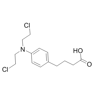 pools of structures, where the assumed flexible parts of the protein are in random conformations. The selected pool of structures does not describe the actual combination of specific structures that is found in the solution, but rather a collection of structures representative of the solution state in size and conformation. As a complementary approach to the SASRFMX and EOM analysis we also employed the program OLIGOMER to evaluate the quaternary structure distribution. From a predefined set of structures of different size and shape, the program calculates a linear combination of the corresponding scattering curves, in accordance with the experimental data. This hence yields the volume fractions of each oligomeric species.
Month: July 2019
Binding and subsequent translocation to the nucleus we observed a potentiation of induction of COX-2 protein
Signifying that some AhR activity is necessary to keep inflammatory protein levels under control. It would not be unreasonable to assume that endogenous AhR ligands present in organs such as the lung maintain constitutive AhR activity at levels that do not cause alterations in gene expression but are sufficient to prevent an exaggerated inflammatory response. Such selective modulation of AhR activity could be why activation of the AhR by CSE repressed COX-2 expression, whereas classic AhR ligands such as TCDD increase COX-2 protein levels. Both CSE and BP increased AhR activation in lung fibroblasts, as evaluated by Cyp1a1 mRNA induction, suggesting that the incongruity of results between CSE and BP is not due to inability of CSE to activate the AhR. Discrepancy in physiological responses to AhR ligands have been observed elsewhere, including murine models of multiple sclerosis. Here, activation of the AhR by either TCDD or ITE suppresses experimental autoimmune encephalomyelitis yet is enhanced by FICZ. Moreover, both ITE and TCDD elicit the same pattern of AhR-dependent gene expression, yet ITE does not cause toxicological outcomes associated with dioxin exposure, suggesting that their divergent mechanisms of action may be independent of classic AhR activation. The AhR binds to a structurally-diverse array of ligands, and it has been postulated that differential binding to the AhR may contribute to divergence in overall functionality. The high plasticity of ligand effects on signaling pathways, including ligand-dependent differences in co-factor recruitment to target genes, may account for some of this variance. Our results show that suppression of COX-2 protein in response to CSE is due to the AhR, concurrent with HuR nuclear retention, both of which did not occur with classic AhR ligands, suggests divergent mechanisms of COX-2 regulation by the AhR. Despite our results showing lack of Cox-2 mRNA induction in absence of AhR expression, there is a profound increase in COX-2 protein, implicating post-transcriptional mechanisms as the way in which the AhR prevents COX-2 expression. Mammalian cells have evolved post-transcriptional mechanisms that further control inflammatory protein levels. Posttranscriptional control is accomplished by regulating nuclear export, cytoplasmic localization, translation initiation and mRNA decay, the latter being determined by the presence of the ARE in the 39-UTR of mature mRNA. Many transiently-expresses cytokines, growth factors and other mediators, including Cox-2, contain AREs and whose mRNA is rapidly destabilized. Our results demonstrate that CSE-induction of Cox2 mRNA in AhR-expressing cells was transient, and returned to baseline by 6 hours, indicative of rapid mRNA decay. Using ActD, an inhibitor of RNA synthesis, we show that when the AhR is expressed, Cox-2 mRNA is rapidly degraded. Thus, post-transcriptional regulation of protein expression by the AhR may be an important adaptive mechanism to control cellular perturbations caused by environmental stress. Stress responses can profoundly affect 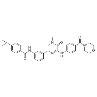 mRNA stability via the concerted efforts of numerous RNA-binding proteins including CUGBP2, TTP and HuR, all of which can play a role in regulating Cox-2 expression. Both GW786034 CUGBP2 and HuR are nuclear proteins, undergoing translocation to the cytoplasm in response to a variety of stress conditions, including c-irradiation, reactive oxygen species and ATP depletion. This nuclear-cytoplasmic shuttling is believed to provide protection against Cox-2 mRNA CX-4945 degradation. The transcriptional regulation of HuR is virtually unknown and there is no information on whether cigarette smoke alters the expression or localization of RNA-binding proteins, including CUGBP2 and HuR. Therefore, our data are the first to show that neither cigarette smoke nor AhR expression.
Profiles of BMAL2 proteins levels were compared in the livers of Wistar rats and SHR subjected to RF
The data revealed that both strains differed in timing of the maximal BMAL2 levels. Our results demonstrate that SHR is behaviorally more sensitive to situations in which food availability is restricted to an improper time of day, developing earlier and stronger FAA than control Wistar rats. Whereas the restriction of food availability phase-shifted the nocturnal locomotor activity of the SHR, it only temporally redistributed this activity in the Wistar rats. The RF-induced behavioral phase shift in the SHR was not mediated by the SCN. Whereas RF exerted similar effects on the colon in both rat strains, it affected the hepatic clock of the SHR differently when compared with the Wistar rats. In the liver of the SHR, the expression profiles of Bmal1 and Bmal2 were synchronously advanced in response to RF, whereas in the Wistar rats only Bmal1 expression was advanced. RF exerted a gene-, tissue- and strain-specific impact on the temporal regulation of the expression of genes that are either driven by the clock or whose protein products enable communication between the clock and metabolism. Altogether, these data suggest that the sensitivity of the circadian system to changes in the timing of food availability may greatly differ between the two closely related rat strains. Moreover, the results suggest a possible role of the Bmal2 gene in the higher sensitivity of the hepatic clock of SHR to RF. SHR/Ola exhibit a lean phenotype with body weight typically lower compared with Wistar rats. Under ad libitum feeding, SHR spent more time eating Silmitasertib during the nighttime than during the daytime. The percentage of time spent eating during the day seems to be higher than what we found previously in Wistar rats, which was in agreement with the findings of others. This was also in agreement with previous observations of diminished diurnal variation in the feeding behavior of SHR compared with WKY rats as a result of increased food intake during the light period and mild decreases during dark periods. It is possible that our result relates to our previous observation of a positive phase-angle of entrainment of the SCN-driven rhythms in SHR maintained on a LD cycle. The rats became active prior to the onset of darkness and thus likely consumed more food during the daytime. During exposure to RF, the rats were allowed to consume food for only 6 h per day, which imposed a significant daily rhythm in the food intake of the animals. Moreover, the food was made available at improper times of day to increase the urgency of this signal. Nevertheless, the 6-h feeding regime likely represented sufficiently long interval to maintain normocaloric intake Epoxomicin 134381-21-8 because rats of both strains gained weight during the 10 days of RF. Interestingly, under RF SHR consumed significantly more food and gained slightly more weight relative to their body weight than Wistar rats. Therefore, the observed increase in sensitivity to RF in SHR was not due to a change in caloric intake on RF. Our data demonstrate that the exposure of rats to RF induces FAA in both rat strains. However, the FAA occurred much earlier and was more pronounced in the SHR than in the Wistar rats, suggesting that for SHR, RF may present a stronger signal to entrain the putative food entrainable oscillator that drives FAA. In 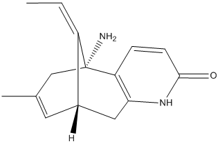 accordance with the finding of stronger FAA in SHR, the locomotor activity under RF was completely phase-advanced according to the time of food availability. In contrast, the locomotor activity of the Wistar rats was redistributed due to RF into only two bouts, one related to food presence and the second remaining synchronized with the external LD cycle. A similar effect of RF on the locomotor activity of Wistar rats has been observed previously elsewhere. The results suggest that whereas in Wistar rats the behavioral activity is entrained by RF as well as by the external LD conditions.
The feeding rhythm is one of the signals that provide temporal information upcoming changes in the external cycle
In mammals, rhythms in feeding and activity represent outputs of the so called “central” clock located in the hypothalamic area of the brain in the suprachiasmatic nuclei. The SCN are specialized to perceive information about external LD cycle due to their direct connection with the retina. The SCN clock adjusts its rhythmicity according to lighting information and passes the signal to so-called “peripheral” clocks, which are located in various bodily cells and drive rhythmically the tissue- and organ-specific programs. As a result, the processes driven by the central and peripheral clocks are synchronized. At the cellular level, the molecular mechanism responsible for generating circadian oscillations is composed of interconnected transcriptional/translational feedback loops among the clock genes and their Tulathromycin B protein products. The core clockwork consists of the transcriptional activators CLOCK and BMAL1, which bind as a heterodimer to E-box elements in the promoters of the clock genes Per1, Per2, Cry1, Cry2, Rev-erb��/�� and Ror��/��/�� and drive their transcription. The PER1,2 and CRY1,2 proteins accumulate in the cytoplasm, form complexes and translocate into the nucleus, where they inhibit the transcriptional activity of CLOCK-BMAL1. This feedback mechanism results in the rhythmic expression of the clock genes. Furthermore, REVERB�� and ROR�� create accessory feedback loops by binding to the promoter in Bmal1 to repress or activate its transcription, respectively. Bmal2 is a paralog of Bmal1, and its expression from a constitutively expressed promoter can restore the clock and metabolic phenotypes of Bmal1-knockout mice. The ROR��-mediated activation of Bmal1 transcription is enhanced by the PPAR�� coactivator 1��. Members of the PAR bZIP transcription factor family, including the activator DBP and the repressor E4BP4, act via Dbox elements in their target genes to form additional accessory feedback loops. Posttranslational modifications, such as phosphorylation, ubiquitination, sumoylation, and acetylation/ deacetylation, enhance the robustness and stability of the clock mechanism. The rhythmic regulation of Ebox elements by the BMAL1/CLOCK complex, D-box elements by PAR bZIP factors, or RORE sequences by ROR��/REVERB��, drive the rhythmic expression of so-called clockcontrolled genes, coding transcriptional factors or functional proteins involved in the regulation of various physiological pathways. In nature, food is rarely present ad libitum, and feeding regimes are affected by many factors. Under laboratory conditions, limiting food Folinic acid calcium salt pentahydrate access to a certain time of day, socalled restricted feeding, leads to the development of food-anticipatory behavior in advance of food availability, including increases in locomotor activity, body temperature, corticosterone levels, and plasma levels of ketone body and free fatty acids. Food-anticipatory activity is driven independently of the central clock because it occurs in SCN-lesioned animals as well as in animals with a genetically ablated clock. This phenomenon is thought to be driven by another oscillator, the so called food-entrainable clock, as it persists in conditions without periodic food availability. The food-entrainable oscillator likely communicates with the circadian clock, as FAA is absent when the period of the feeding cycle is too distant from 24 h. Moreover, this SCN-independent oscillator may depend partially on the canonical circadian clock components, as deficiencies in individual clock genes lead to variations in the circadian range of FAA entrainment. Therefore, clock genes are 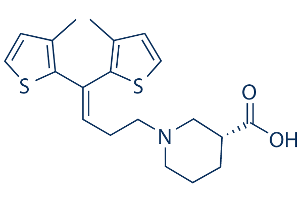 likely involved in the regulation of FAA in a circadian oscillatory manner. However, in spite of significant efforts to determine its localization in the body, the oscillator has not yet been identified. The SCN pathways entraining the peripheral clocks are of multiple origin.
J82 cells react with decreased proliferation to inhibition of HA synthesis achieved either by knock-down
Moreover, high expression of Hyal1 has recently been suggested to predict progressive muscle-invasive and recurrent BC and to associate with metastasis and decreased disease specific survival. All together, the available data on HA in BC point to increased synthesis of HA mainly by HAS1 and degradation to tumor promoting sHA by Hyal 1. In turn, the effects of HA and sHA are likely transduced through activation of HA-receptors. In BC certain splice variants of CD44, such as CD44V8-10, were Butenafine hydrochloride upregulated and associated with poor prognosis. In addition, it has been shown that CD44 might be involved in the angiogenic response to HA in BC. However, a recent analysis revealed that CD44v was upregulated and CD44s was downregulated without diagnostic or prognostic value. RHAMM has been described to be upregulated but also without clear correlation with clinical parameters. Because it appears plausible that the amount of HA-receptors plays an important role in the transduction of HA signaling and thereby in the progression of human BC, the aim of the present study was to study the expression of HA-receptors and HAS isoenzymes and to achieve mechanistic information in a patient collective characterized by >50% muscle-invasive BC. Patients suffering from urothelial transitional cell cancer of the bclinical outcometheir prognosis. Patients with non-muscleinvasive BC are treated by TUR of the tumor. In most cases this procedure combines diagnostic and curative treatment leading to excellent tumor control. Within TUR representative tissue samples can be taken for histopathological diagnosis and determine the necessity of more aggressive treatment. For many patients this local procedure can become a long-term bladder preserving strategy. However, 3-5% of the patients suffering from nonmuscle-invasive BC experience progression to muscle-invasive disease and need radical treatment. Only about 50% of the patients with muscle-invasive BC benefit from the radical operation with long-term disease-free survival. To  develop more individualized therapeutic strategies, it is crucial to identify tumor-related parameters allowing risk stratification within the group of muscle-infiltrating transitional cell carcinomas and in the long run represent potential therapeutic targets. In this regard HA and HA-associated genes, such as HAS isoenzymes, HA receptors and HA catabolic enzymes, appear to be interesting because these genes are of pathophysiological significance and of diagnostic value in various cancer entities. Interestingly, the association of HA with cancer parenchyma or with tumor stroma is specific for certain cancer types and can also be stage-dependent. These observations suggest that HA affects both tumor cells and stromal cells. Furthermore, it is likely that tumor-associated fibroblasts can LOUREIRIN-B secrete HA that is subsequently utilized by tumor cells. The expression of HA-related genes has been studied also in BC. As a result, especially HAS1, Hyal1 and urinary HA excretion were found to be associated with BC progression and/or prognosis. The present investigation revealed a stage-dependent increase of RHAMM expression in human BC specimens as evidenced by real-time RT PCR and by immunohistochemistry. Of note, high RHAMM expression levels were associated with increased disease-specific and increased overall mortality. Importantly, in this study we present for the first time that RHAMM mRNA expression is even an independent risk factor in muscle-invasive BC. Subsequently knock-down of RHAMM caused significant inhibition of xenograft tumor growth in nu/nu athymic mice. This reduction of tumor progression was likely caused by inhibited proliferation of BC tumor cells as evidenced by Ki67 staining. Further in vitro experiments verified the antiproliferative effect of RHAMM knock-down.