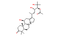
This kinetic analysis confirmed that myoA KO Benzoylaconine parasites moved slowly for a short distance, and usually stopped after,14 mm for several minutes, before forming another semicircle, which confirmed the results of the trail deposition assay. Next, we compared invasion and replication rates between myoA KO and WT parasites.  First, parasites were allowed to invade HFF cells for different times and left to replicate for 24 hours. Since one of the earliest markers of the entry process is the TJ, it was important to assess if the observed reduction in invasion rate was caused by a delay in TJ formation, or by a block in parasite progression into the host cell after TJ formation. While myoA KO parasites invaded the cell via a normally RON-shaped TJ that the majority of myoA KO parasites remained attached to the host cell without forming a TJ. Approximately 30% of myoA KO parasites formed a TJ and another 10% of all parasites were internalized. In contrast, the majority of control parasites were found to be intracellular, 10% were in the process of entry and only 10% still remained extracellular without TJ initiation. Together these data demonstrate that a step upstream of TJ formation, such as host cell recognition or reorientation of the parasites is delayed in absence of MyoA. To directly analyse the effect of MyoA depletion on host cell entry, the kinetics of this invasion step was analysed using timelapse microscopy. In total 22 entry events for the control and 27 for myoA KO parasites were compared. As expected, control parasites moved in within 20�C30 seconds in a smooth and uniform movement. In contrast, myoA KO parasites showed huge variability, with some parasites penetrating the host cell rapidly in a smooth process. However, the majority 4-(Benzyloxy)phenol entered in a spasmodic stop-and-go fashion, and appeared stalled for several seconds to minutes. The fastest recorded entry was,25 seconds, whereas the slowest entry took almost 10 minutes until the parasite was completely internalised. While these results demonstrate an important function of MyoA for efficient, smooth host cell penetration, the fact that myoA KO parasites remain capable of penetrating at a similar speed to wild-type parasites indicates that the force for host cell penetration can be generated independently of MyoA. Together these results were interpreted to be that MyoA plays an important but not essential function in multiple steps during host cell invasion. Since deletion of other components of the invasion machinery impeded mutant survival, we speculated that the function of MyoA might be partially complemented by other myosins. Together these data demonstrate that GAP45 has a role in providing the IMC with its typical structure, thereby promoting the anchorage of the MyoA motor complex in the IMC. However, since gliding motility was less affected, it appears that the parasite can efficiently produce forward movement in the absence of a myosin motor that is properly anchored along the IMC. In addition, the significant loss of invasiveness is unlikely to result from an impairment of gliding motility as previously suggested, but is rather caused by the morphological defects of these mutants. While the study of the motor mutants demonstrates that alternative pathways must be in place that can drive gliding motility and invasion, it does not rule out a critical function of other myosin motors.